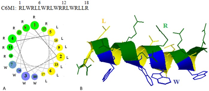
Figure 1. Helical structure and helical wheels representation of C6M1.

A) A downward cross-sectional view of the helix axis is shown. The axis of the alpha helix is orthogonal to the paper plane. The bigger the circle is, the closer is the location of the residue to the upper end, when viewing from the top, B) In helical structure, same amino acids (side chains) face the same side of the helix. The schematics was generated using RaptorX web server [24]. R (green), L (yellow), and W (blue) represent arginine, leucine and tryptophan residues, respectively.
