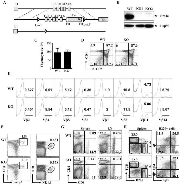
Figure 2. Deficiency of Itm2a has little impact on the differentiation of immune cells.
A. Generation of Itm2a-deficient mice. Schematic diagrams of Itm2a gene (top), the targeting construct (middle), and deleted allele (bottom) are shown. Exons are represented with gray boxes and numbered. Regions for homologous recombination are marked with dashed lines. Neo and TK stand for neomycin cassette and thymidine kinase. B. T cells were purified from control (WT) and two Itm2aKO mice and stimulated with anti-CD3 for 72 hours. Cell extract from the stimulated cells was probed with anti-Itm2a and anti-Hsp90. C. Thymocytes of WT and Itm2aKO mice were enumerated, and the means and standard deviations are shown in the left panel. D & E. The thymocytes were also stained with anti-CD4/anti-CD8 (D) and anti- α/β TCR, and TCRhigh cells were further stained with antibodies against various Vβ chains indicated in E. F. The thymocytes were stained with anti-CD4/anti-Foxp3 and anti-TCR/NK1.1. G. Splenocytes and lymph node cells of WT and Itm2aKO mice were stained with anti-CD4 and anti-CD8. H. The splenocytes were also stained with anti-TCR/anti-B220. B220+ cells were then further separated by anti-IgM/anti-IgD staining. The data shown in D–H are representative FACS plots of at least three independent experiments.

