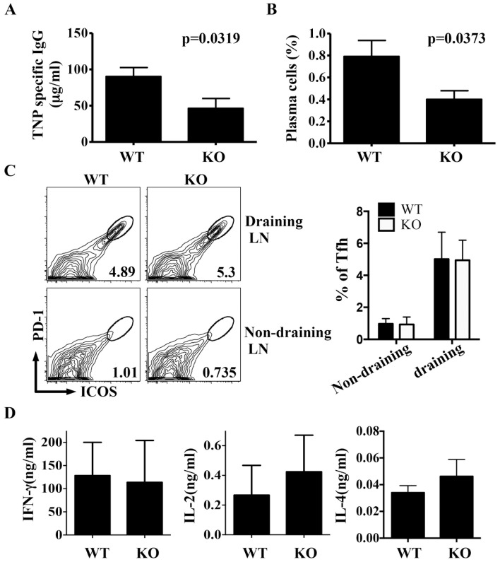
Figure 6. Impaired immune response to Th cell-dependent antigens in the absence of Itm2a.
WT (N = 8) and Itm2aKO (N = 7) mice were immunized with TNP-KLH in CFA at tail base. Two weeks after the immunization, the level of TNP-specific IgG was quantified with ELISA (A). The percentage of plasma cells (CD138+B220dull) among splenocytes was calculated (B). T cells of draining (inguinal) and non-draining (axillary) lymph nodes were further stained with anti-ICOS/anti-PD-1. ICOS+PD-1+ Tfh cells were gated (C). The means and standard deviations of the percentage of Tfh among lymph node Th cells are shown in the right panel of C. Draining LN cells were restimulated with TNP-KLH for 24 hours. The concentration of IFN-γ and IL-2 in the supernatant of the stimulated cells was quantified with ELISA (D). Statistical analyses were performed with unpaired Student's t tests.

