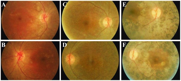
Figure 3. Fundus photographs of the included patients.
(A–B) Fundus photographs of the 24-year-old proband of family USH01 reveal attenuation of retinal vessels, pallor of optic disc, and pigmentary migration, indicating an atypical RP fundus. Macular region is not affected in both eyes. (C–D) Fundus appearance of patient USH01-III2 (22-year-old) is similar to that of patient USH01-III1. Attenuated retinal vessels and pale optic disc are observed. Macular region is also preserved. (E–F) Typical RP fundus is shown by patient USH03-II1, including bone spicule-like pigmentation, retinal vascular attenuation, pallor of optic disk, and chorioretinal atrophy. Macular degeneration is observed in this patient.

