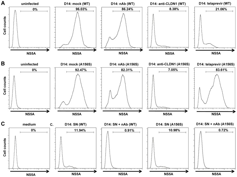
Figure 3. Cell-cell transmission is the main transmission route for DAA-resistant viruses.
The spread assay was performed as shown in Figure 2 with or without 25 µg/mL anti-HCV IgG to neutralize cell-free transmission of virus. The relative percentage HCV-positive cells/total cells was determined by immunostaining for NS5A and flow cytometry. Huh7.5.1 uninfected cells were used as a negative control (“uninfected”). (A–B) Percentage of wild-type (WT) (A) or DAA-resistant HCV (A156S) (B)-infected cells without treatment (mock) or in the presence of 25 µg/mL anti-HCV IgG (nAb), 10 µg/mL anti-CLDN1 mAb or 1 µM telaprevir treatment as described in Figure 2 is shown. (C) The supernatant (SN) with or without anti-HCV IgG containing cell-free wild-type or A156S HCV was used to infect fresh Huh7.5.1 cells. The cell culture medium was taken as a negative control. The data are represented of three experiments.

