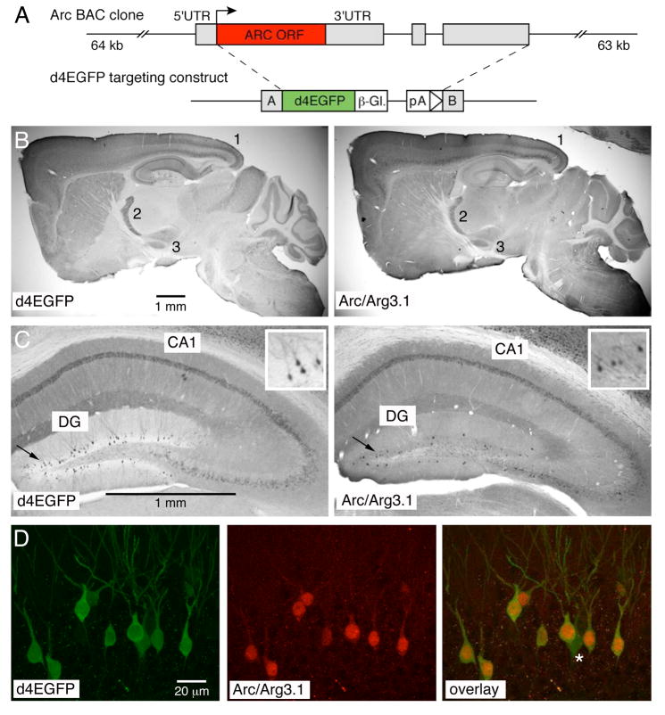
Figure 1. Comparison of d4EGFP and native Arc/Arg3.1 expression.
(A) Top: schema of the 127 kb BAC clone; Arc/Arg3.1 coding sequence is shown as a red box, UTR’s as gray boxes. Bottom: d4EGFP construct used to generate the BAC-derived TgArc/Arg3.1-d4EGFP transgene; d4EGFP coding sequence is shown as a green box, β-globin poly-A and intron as an interrupted white box, and the targeting Arms A and B as gray boxes. (B) Immunohistochemistry with anti-GFP (left panel) and anti-Arc/Arg3.1 antibodies (right panel) reveals overall comparable patter of expression, with prominent labeling in deep and superficial cortical layers (1), reticular thalamus (2) and subthalamic nucleus (3); the immunosignal was visualized with HRP-conjugated secondary antibodies and DAB reaction; see also Supplemental Figure 1 for comparison of all nine transgenic lines and Arc/Arg3.1 mRNA expression. (C) Higher magnification of anti-GFP (left panel) and anti-Arc/Arg3.1 immunosignal in the hippocampus. Note the prominent labeling of many cells in the CA1 and sparse labeling in the dentate gyrus (DG) regions, respectively. Insert in the right upper corner of each panel shows zoom-in view of the DG labeling. (D) Confocal microscopy of direct d4EGFP signal (left panel) and Cy3-visualized anti-Arc/Arg3.1 immunosignal (mid panel) reveals high level of colocalization in the DG region (overlay, right panel). See Supplemental Figure 2 for further examples of d4EGFP and Arc/Arg3.1 colocalization in the DG and cortex.

