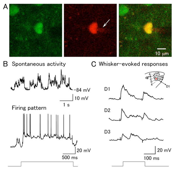
Figure 5. In vivo whole-cell recording of d4EGFP-positive neurons in the barrel cortex.
(A) An example of a d4EGFP-positive in vivo-patched layer 2/3 neuron. Left: d4EGFP signal; middle: Alexa 594 red signal in the internal recording solution, with a visable shadow of the patch pipette tip (white arrow); right overlay. (B) Spontaneous activity and firing pattern of d4EGFP-positive neuron. Top trace shows typical two-state membrane fluctuations (up and down states) observed in cortical neurons in anesthetized animals; bottom trace shows spiking in the same cell, elicited with DC current injection. (C) Deflection of the D1, D2, and D3 whiskers, as illustrated in the schema on the top right, evoked the corresponding responses shown below. The time course of the whisker deflection is shown at the bottom.

