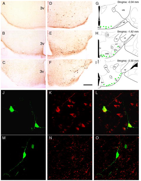
Figure 7. Induction of d4EGFP in the arcuate nucleus and retrochiasmatic area in immune stress.
Immunohistochemistry with anti-GFP antibody revealed no signal in control mice (A–C) and a profound upregulation of d4EGFP expression 6 hrs after a single LPS injection in the ventral hypothalamus (D–F). d4EGFP-positive neurons were mainly located in the lateral part of the arcuate nucleus and retrochiasmatic nucleus (extending to the lateral tuberal region), as represented by green circles in the schematic drawings in G–I (Paxinos, Franklin, 2003). Co-staining with antibodies against EGFP (J, M), beta-endorphin (K) or neuropeptide Y (N) revealed overlap of d4EGFP expression with β-endorphin (L) but not NPY (O). Scale bars = 200 μm (A–F), 40 μm (J–O). Abbreviations: 3v - third ventricle, Arc - arcuate nucleus, cp - cerebral peduncle, f - fornix, LH - lateral hypothalamus; mfb - medial forebrain bundle, mt - mammillothalamic tract, MTu - medial tuberal nucleus, ns - nigrostriatal bundle, opt - optic tract, RCh - retrochiasmatic area, Te - Terete hypothalamic nucleus, VMH - ventromedial hypothalamic nucleus.

