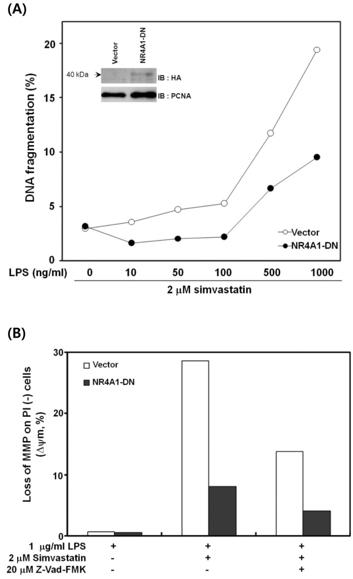
Figure 3.
The effect of dominant-negative NR4A1 expression on apoptosis. (A) RAW 264.7 cells were transfected with control or dominant-negative NR4A1 (NR4A1-DN) vectors and treated with various concentrations of LPS and 2µM simvastatin for 12 h. Using flow cytometry, the percentage of subgenomic DNA content was measured in propidium iodide (PI)-stained cells to evaluate DNA fragmentation. The inset shows the presence of NR4A1-DN in the nucleus of transfected cells. (B) RAW 264.7 cells transfected with control or NR4A1-DN vectors were incubated in LPS or simvastatin alone or in combination with Z-VAD-FMK for 12 h and stained using rhodamine-123. The mitochondrial membrane potential (MMP) of the stained cells was analyzed using flow cytometry. A representative result of four experiments is shown.

