FIGURE 4.

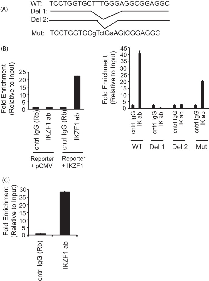
Ikaros is recruited to the specific site in the intron of PP2A. A, schematic of the mutant (Mut) reporters used in these experiments. B, reporter ChIP in 293T cells. 293T cells were transfected with either only the wild type (left panel) or the mutant reporter (right panel) or the reporter in combination with the Ikaros expression plasmid using Lipofectamine 2000. Twenty-four hours after transfection, cells were collected, and a ChIP assay was performed using the MAGnify ChIP kit. The region spanning the specific intron site was amplified by quantitative PCR and normalized to the values obtained from the input DNA. The graph shows mean ± S.D. of three observations. cntrl, control; ab, antibody; Rb, rabbit. C, ChIP assay in primary T cells. 5 million freshly isolated primary T cells were used for each antibody/sample. There was no transfection, and endogenous proteins were used for immunoprecipitation of the chromatin. A ChIP assay was carried out using the MAGnify ChIP kit. The region spanning the specific intron site was amplified by quantitative PCR and normalized to the values obtained from the input DNA. The graph shows mean ± S.D. of three observations.
