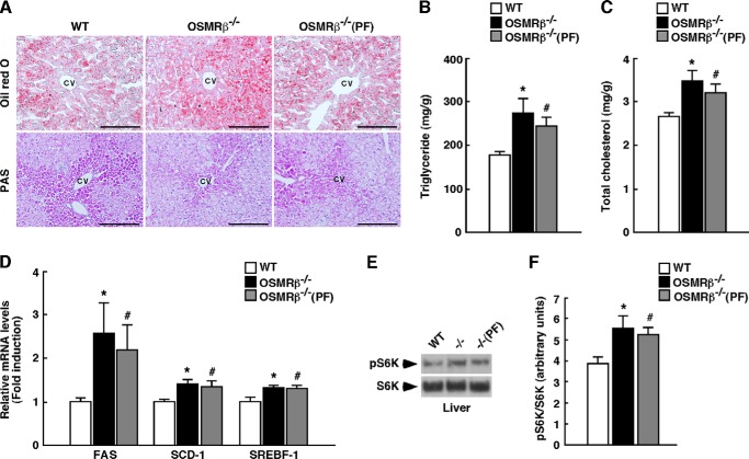
FIGURE 4.
Lipid metabolism in the livers of WT and OSMRβ−/− mice under HFD conditions. The mice (8 weeks old) were fed an HFD for 8 weeks. A, Oil Red O and PAS staining of the livers of WT, OSMRβ−/−, and OSMRβ−/−(PF) mice. CV, central vein. Scale bar = 100 μm. B and C, the content of triglycerides (B) and total cholesterol (C) in the livers of WT, OSMRβ−/−, and OSMRβ−/−(PF) mice in the fed state (n = 6). D, the expression levels of genes related to fatty acid synthesis (FAS, SCD-1, and SREBF-1) in the livers of WT, OSMRβ−/−, and OSMRβ−/−(PF) mice in the fed state (n = 6). E, Western blot analysis of phosphorylation of S6K (pS6K) in the livers of WT, OSMRβ−/−, and OSMRβ−/−(PF) mice in the fed state. F, a quantitative analysis of pS6K (n = 6). In the fed state, mice were fasted for 4 h before the experiments to eliminate the feeding effects on lipid metabolism. The data represent the mean ± S.E. *, p < 0.05 WT versus OSMRβ−/− mice; #, p < 0.05 WT versus OSMRβ−/−(PF) mice, Student's t test.

