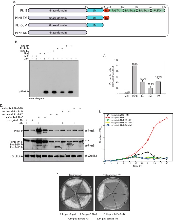
FIGURE 4.
Extracellular domain of PknB is essential for in vitro growth. A, schematic representation of PknB and deletion constructs. B, in vitro kinase assays performed using 80 nm MBP or PknB or PknB deletion mutants, 3.33 μm GarA, 10 μm ATP, and 10 μCi of [γ-32P]ATP. Samples were resolved on 12% SDS-PAGE and autoradiographed. C, bands corresponding to phosphorylated GarA (p-GarA) were excised from the gel and quantified by liquid scintillation counting in three independent experiments. Activity of PknB was normalized to 100% in each experiment, and the percentage of activity in other samples was calculated with respect to (w.r.t.) PknB activity. The results were plotted with percentage of activity on the y axis and the samples on the x axis in the form of histograms. D, cultures were seeded at an initial A600 of 0.2. Cultures were grown in the presence or absence of ATc as indicated above for 6 h. For mc2ΔpknB::pNit, mc2ΔpknB::PknB-KD, mc2ΔpknB::PknB-JM, and mc2ΔpknB::PknB-TM strains, 0.2 μm IVN inducer was added during depletion. WCLs prepared were subjected to Western blotting with α-PknB and α-GroEL1 antibodies. The arrows indicate bands corresponding to PknB deletion mutants. E, growth pattern analysis of mc2ΔpknB transformants grown in the absence of ATc. Cultures were seeded at an initial A600 of 0.02. mc2ΔpknB::PknB-KD, mc2ΔpknB::PknB-JM, and mc2ΔpknB::PknB-TM were grown in the absence of ATc and in the presence of 0.2 μm inducer IVN. F, the Rv-pptr-B transformant strains were streaked on 7H10-agar plates containing 2 μg/ml pristinamycin or 0.2 μm IVN. Error bars, S.E.

