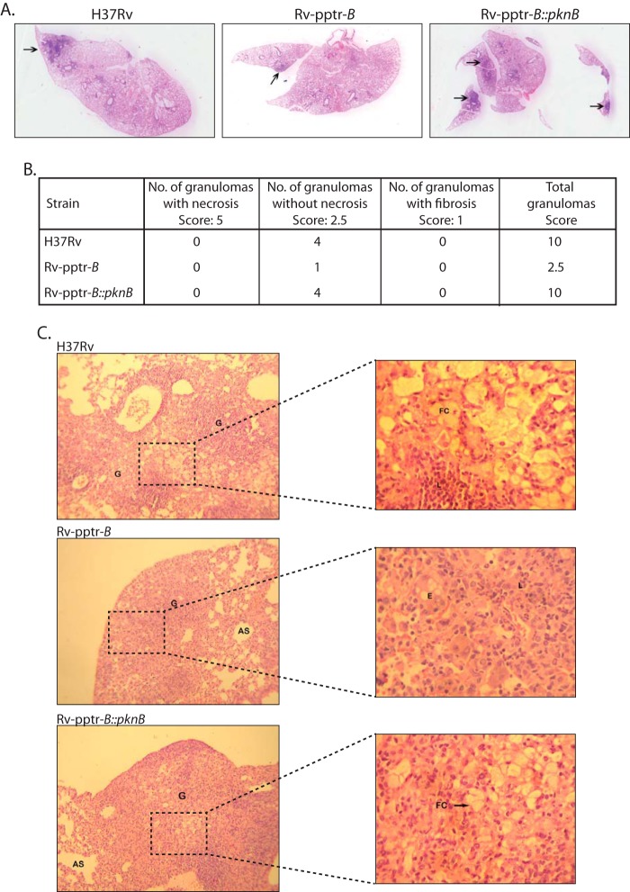
FIGURE 9.
PknB plays indispensable role in the pathogenesis. A, histopathological examination of six sections from each candidate mouse was carried out. The images shown are representative of typical lung sections in each experimental group. B, granuloma score of H&E-stained lung sections for each experimental group was performed as described. C, images depict H&E-stained lung sections of mice infected with H37Rv or Rv-pptr-B or Rv-pptr-B::PknB after 8 weeks. Images at ×100 are shown in the left panels. Images of boxed regions at ×400 are depicted in the corresponding right panels. G, granuloma; L, lymphocytes; FC, foamy histiocytic cells; AS, alveolar spaces; E, epithelioid cells.

