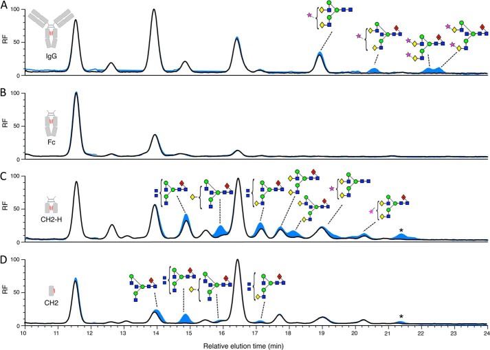
FIGURE 3.
Analysis of EndoS-released glycans from serum IgG and IgG fragments. HPLC analysis of fluorescently labeled glycans released from serum IgG (A) and recombinantly expressed Fc (B), CH2-H (C), and CH2 (D) using EndoS (black) and sequential digest with EndoS then PNGase F (blue). Peaks were assigned using negative ion electrospray ionization-MS/MS. Glycans that remained after digest with EndoS, which were then cleaved as a result of sequential digest with PNGase F, are labeled using Oxford glycan nomenclature (46). The star denotes glycan with the composition Neu5Ac2Hex5GlcNAc4 (probably Neu5Ac2Gal2GlcNAc2Man3GlcNAc2) that was present in too low a quantity for fragmentation analysis. Fluorescence is reported as relative fluorescence units (RF).

