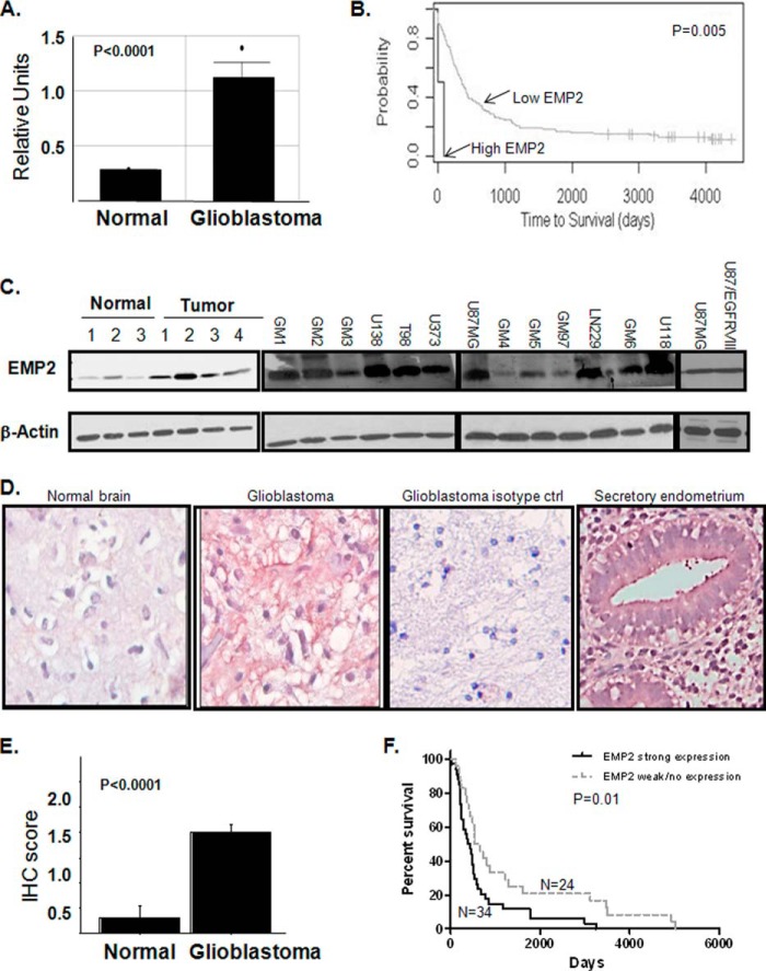
FIGURE 1.
EMP2 expression is increased in GBM. A, EMP2 mRNA expression (Affymetrix microarray) was increased in GBM compared with normal brain. *, p < 0.001. B, survival data for high and low EMP2 mRNA expression. C, left, EMP2 expression was increased in GBM tumors compared with normal regions. Right, EMP2 expression was evaluated in a panel of GBM cells lines and in lines derived from patients by Western blotting analysis. β-Actin expression was used as a loading control. D and E, GBM tissue arrays containing 329 cores from 110 patients were stained for EMP2 expression. D, EMP2 protein expression was determined in normal brain, in GBM, and in secretory endometrium (positive control) using an EMP2 polyclonal antibody. To detail nonspecific staining, rabbit preimmune serum was used. Staining was visualized using deNovo Red. Nuclei were counter-stained using hematoxylin. E, EMP2 expression was quantitated on a 0–3 histological scale by two independent pathologists, and the average IHC score is shown. F, EMP2 expression was dichotomized based on high (histological score, ≥2) or low (histological score, ≥1) expression. High EMP2 expression correlated with a poor survival.

