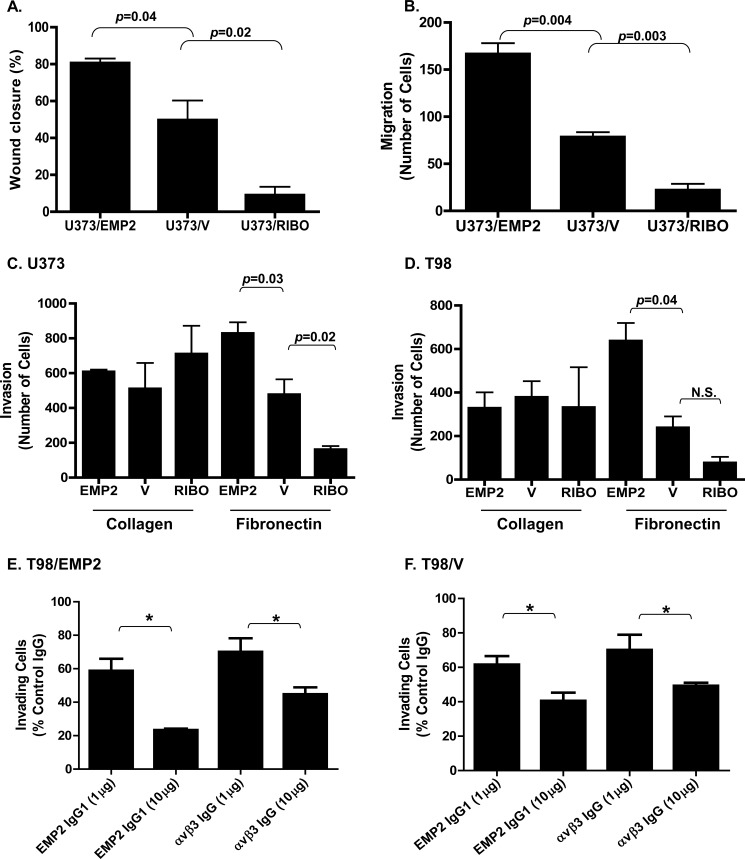
FIGURE 3.
EMP2 promotes GBM invasion. A, U373/EMP2, U373/V, or U373/RIBO cells were grown to form a monolayer. A scratch was then created, and closure of the wound was measured after 24 h. Experiments were performed at least three times, and the results were averaged. B, equal numbers of U373/EMP2, U373/V, and U373/RIBO were plated into the top of the transwell. After 6 h, cells that had migrated through the transwell were fixed, stained with crystal violent, and counted. Values are averages of three independent experiments (±S.E.). U373/EMP2, U373/V, or U373/RIBO cells (C) or T98/EMP2, T98/V, or T98/RIBO cells (D) were added to transwells coated with collagen I or fibronectin. Cells that had invaded through the transwell were determined as above. The experiment was repeated three times, with the data presented as the mean ± S.E. A–D, Student's t test was used to determine significant differences between groups with specific p values indicated in the figure; N.S., not significant. T98/EMP2 (E) or T98/V (F) cells were preincubated with varying concentrations of an anti-EMP2 IgG1, anti-αvβ3 integrin, or isotype control antibody. Cells were then plated onto a fibronectin-precoated transwell, and percent invasion relative to the isotype control was determined. Results represent averaged results from three independent experiments ± S.E. *, p < 0.05.

