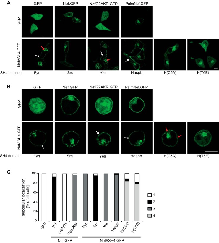
FIGURE 2.
Subcellular localization of chimeric Nef.GFP proteins in transfected cells. A, live cell imaging of HeLa cells transiently expressing the indicated Nef.GFP proteins. Cells were analyzed by confocal microscopy 24 h post-transfection. Individual representative Z sections are shown. Arrows indicate localization at PM (white arrows) or strong intracellular accumulation (red arrows), respectively. B, transfected Jurkat TAg cells were plated on poly-l-lysine-coated cover glasses, fixed 24 h post-transfection, and analyzed by confocal microscopy. Individual representative Z sections are shown. Arrows indicate localization at PM (white arrows) or in the cytoplasm (red arrows), respectively. Scale bar, 10 μm. C, quantification of the frequency of transfected HeLa cells displaying the following subcellular localizations: category 1 (white bars), cytoplasm; category 2 (black bars), cytoplasm, intracellular accumulation, PM; category 3 (dark gray bars), intracellular accumulation, PM; category 4 (light gray bars), cytoplasm, intracellular accumulation. HeLa cells transfected with the different Nef.GFP expression plasmids were grown on cover glasses and fixed 24 h post-transfection. At least 100 cells were analyzed per condition.

