FIGURE 3.

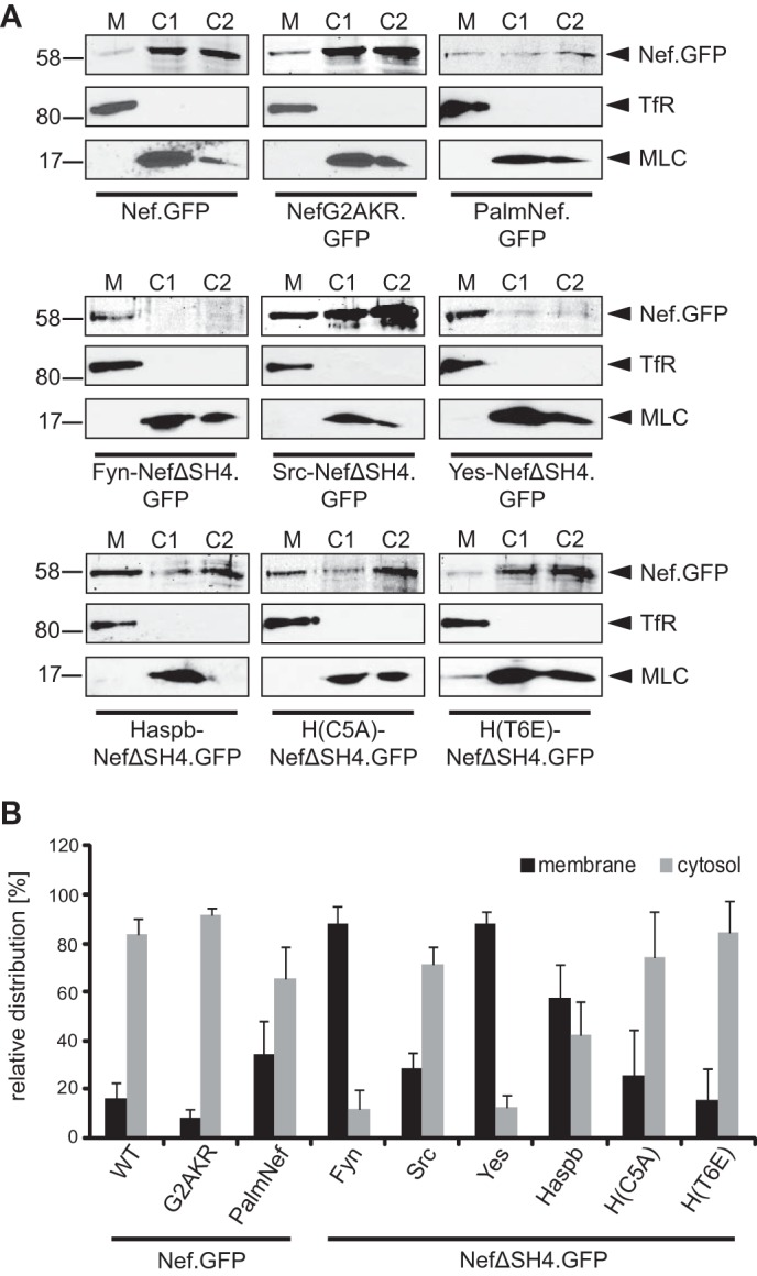
Membrane association of chimeric Nef.GFP proteins in transduced Jurkat TAg cells. A, membrane fractionation of Jurkat TAg cells transduced with lentiviral vectors encoding for the indicated Nef.GFP proteins. Seventy-two hours post-transduction, cell lysates were fractionated into membrane (M) and cytosolic (C1 and C2) fractions and analyzed by Western blotting for the distribution of Nef.GFP. Transferrin receptor (TfR) and myosin light chain (MLC) proteins were analyzed as markers for M and C1/C2 fractions, respectively. B, quantification of the membrane fractionation analysis presented in A. Shown is the relative distribution of the various Nef.GFP proteins into M and C1/C2 fractions with the total signal from both fractions set to 100%. The data represent average values from at least three independent experiments with the indicated standard error of the mean.
