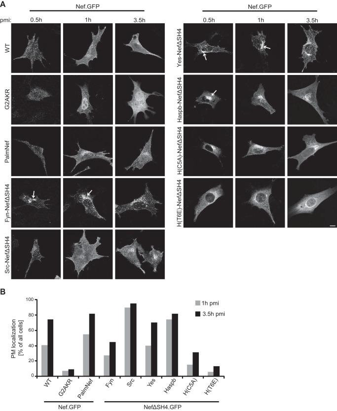
FIGURE 4.
Subcellular distribution of newly synthesized chimeric Nef.GFP proteins. A, NIH 3T3 cells were microinjected with expression plasmids for the different chimeric Nef.GFP proteins and fixed at the indicated time points after microinjection. pmi, postmicroinjection. Cells were analyzed by confocal microscopy. Individual representative Z sections are shown. Arrows indicate different subcellular localization of Fyn-, Yes-, and Haspb-NefΔSH4 compared with Nef WT and Src-NefΔSH4. Scale bar, 10 μm. B, quantification of the frequency of cells displaying PM localization 1 and 3.5 h postmicroinjection of the indicated Nef.GFP proteins. At least 20 cells were analyzed per condition. The 0.5-h postmicroinjection time point was not quantified as low protein expression at this early point precluded thorough analysis.

