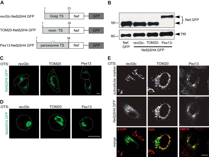
FIGURE 6.
Subcellular localization of Nef.GFP expression constructs containing different organelle targeting signals. A, schematic representation of Nef.GFP constructs used. To achieve organelle specific targeting, the Nef SH4 domain was replaced by the shown targeting signals (TS). mom, mitochondrial outer membrane. B, Western blot analysis of transfected Jurkat TAg cells with the Nef.GFP expression constructs. Equal amounts of GFP positive cells were loaded per lane based on transfection efficiency (%). Transferrin receptor serves as loading control. C, live cell imaging of HeLa cells transiently expressing the indicated Nef.GFP proteins. Twenty-four hours post-transfection, cells were analyzed by confocal microscopy. Individual representative Z sections are shown. Cell circumferences are indicated by gray dotted lines. D, Jurkat TAg cells, transfected with the different Nef.GFP expression plasmids, were plated on poly-l-lysine-coated cover glasses, fixed 24 h post-transfection, and analyzed by confocal microscopy. Individual representative Z sections are shown. Cell circumferences are indicated by gray dotted lines. E, HeLa cells, transfected with the different Nef.GFP expression plasmids, were grown on cover glasses, fixed 24 h post-transfection, and stained for subcellular markers: β-COP (Golgi marker), Cytc (mitochondrial marker), or PMP70 (peroxisome marker), respectively, using fluorescently labeled antibodies. Shown is staining of the indicated subcellular marker (red), GFP (green), and the merge channel. Cells were analyzed by confocal microscopy. Individual representative Z sections are shown. Scale bar, 10 μm.

