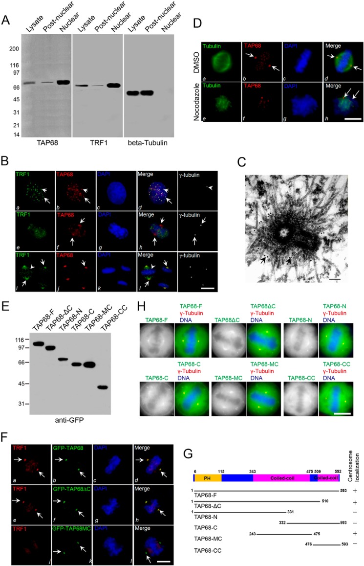
FIGURE 3.
TAP68 mediates TRF1 localization to the centrosome. A, TAP68 and TRF1 share similar subcellular localization. Nuclear and post-nuclear fractions were generated from interphase HeLa cell homogenates. Equal amounts of proteins (35 μg) from various fractions were separated by 5–15% gradient SDS-PAGE and detected by Western blot analyses using antibodies against TAP68, TRF1, and β-tubulin, respectively. B, representative images of interphase (top panels), late prophase/early prometaphase (middle panels), and metaphase (bottom panels) HeLa cells stained for TRF1 (green), TAP68 (red), DAPI (blue), and γ-tubulin (white). In interphase cells, TRF1 (panel a) and TAP68 (panel b) are readily seen in the nucleus as speckles and dots in the merge panel (panel d). In late prophase or the earliest prometaphase cells marked by nuclear membrane fragmentation, TAP68 appears at the centrosome (panel f, arrows), and TRF1 remains as speckles in the partially condensed chromosome (panel e, arrows). Both TAP68 and TRF1 become co-localized to the centrosome as the chromosomes align or after sister chromatid separation (panels i and j, arrows). A merged image shows the co-localization of TRF1 and TAP68 to the centrosome of mitotic cells (panel l). Bars, 10 μm. C, immunoelectron microscopy of late prophase cells indicated that TAP68 localizes to the pericentrioles. D, representative immunofluorescence images of HeLa cells treated with nocodazole or DMSO. 10 h after drug treatment, the cells were fixed and stained for tubulin (green), TAP68 (red), and DAPI (blue). TAP68 remained centrosome-associated in the absence of microtubules (panel h, arrows). E, ectopic expression of different GFP-tagged TAP68 proteins. HeLa cells were transfected GFP-tagged full-length TAP68 or its deletion mutants. After 36 h, HeLa cells were then harvested for Western blot analysis using a GFP antibody. F, representative images collected from metaphase HeLa cells transiently transfected with GFP-tagged TAP68 and its deletion mutants and stained for TRF1 (red), TAP68 (green), and DAPI (blue). In cells expressing full-length TAP68, both TAP68 and TRF1 are co-localized to the centrosome (panels a, b, and d, arrows). A similar co-distribution of TRF1 with TAP68 was observed in TAP68ΔC mutant-expressing cells (panels e, f, and h) but not TAP68MC-expressing cells (panels i, j, and l), demonstrating that the centrosomal localization region of TAP68 is independent of its TRF1-binding domain. Bars, 10 μm. G, schematic representation of the regions of TAP68, which specifies its centrosomal localization. H, representative images of HeLa cells transfected with different GFP-tagged TAP68 constructs. 24 h after transfection, cells were fix and co-stained for γ-tubulin (red) and DNA (blue). Bars, 10 μm.

