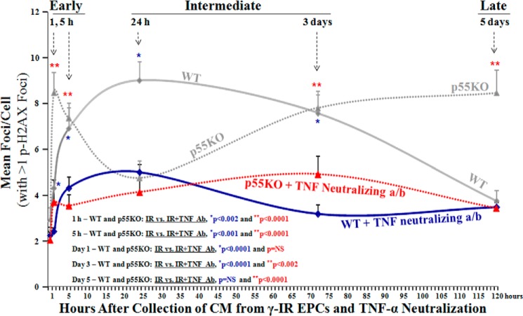
FIGURE 4.
Neutralization of TNF-α in IR-CM resulted in significantly decreased p-H2AX foci formation in TNFR1/p55KO and WT EPCs in vitro. Please note that for clarity of the comparison between the formation p-H2AX foci in EPCs treated with IR-CM media with or without TNF neutralization data points for WT and p55KO, EPCs from Fig. 3 are overplayed in Fig. 4 again. Shown is a graphic representation of mean p-H2AX foci/cell post 24-h treatment of naïve WT and p55KO with IR-CM medium collected and treated with TNF-α neutralizing antibody from respective WT and p55KO EPCs at 1 h, 5 h, 24 h, 3 days, and 5 days post-1-Gy γ-IR. pH2AX foci/cell treated with 1 h IR-CM in WT versus p55KO: 2.4 ± 0.2 versus 3.7 ± 0.6, p < 0.05; pH2AX foci/cell treated with 5 h IR-CM in WT versus p55KO: 4.3 ± 0.5 versus 3.6 ± 0.5, p = NS; pH2AX foci/cell treated with 24 h IR-CM in WT versus p55KO: 5.0 ± 0.5 versus 4.2 ± 0.6, p = NS; pH2AX foci/cell treated with 3-day IR-CM in WT versus p55KO: 3.2 ± 0.4 versus 4.9 ± 0.8, p < 0.03; pH2AX foci/cell treated with 5-day IR-CM in WT versus p55KO: 3.5 ± 0.3 versus 3.5 ± 0.3, p = NS. Treatment of IR-CM with TNF neutralizing antibody decreased the formation of p-H2AX foci at all time points examined in both WT and p55KO EPCs. However, the most significant decreases for WT EPCs were observed at 5 h, 24 h, and 3 days, and those for p55KO EPCs were at 1 h, 5 h, 3 days, and 5 days. Graphs represent data pooled from three independent biological samples treated under similar conditions.

