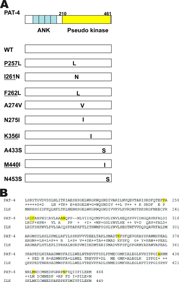
FIGURE 1.
Location of PAT-4 suppressor mutations, effects of these mutations on protein interactions, the sequence alignment of PAT-4 with ILK. A, on the left top is a schematic representation of PAT-4 with ankyrin repeats (ANK) in blue and pseudokinase domain (residues 210–461) in yellow, as defined by PFAM. Below are indicated the nine missense mutations with their approximate locations, all in the pseudokinase domain. Underlining indicates the 5 residues that are identical to those in human ILK. B, ClustalW alignment of the pseudokinase domains of PAT-4 with ILK used to create the homology model of PAT-4. The sequence represented for ILK is the sequence of the protein expressed and crystallized, as reported by Fukuda et al. (24). In the row between the two sequences, identical residues are indicated, and + indicates conservation. Over the span of 266 residues, 59% are identical (157 of 266). Yellow highlighting denotes the residues affected in the nine PAT-4 suppressor mutations.

