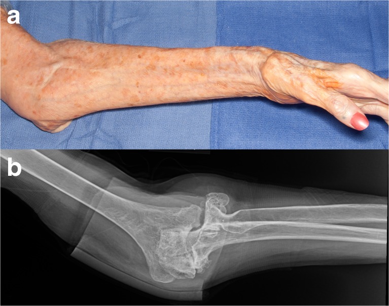
Fig. 1.
Preoperative appearance. a Clinical photograph of the left arm and forearm. The carrying angle was measured at 38°. The left hand shows extensive first dorsal interosseous muscle atrophy. A well-healed scar anterior to the elbow can also be appreciated. b The anteroposterior plain radiograph of the left elbow confirms the extreme cubitus valgus deformity. The head of the left radius cannot be seen. The radial shaft has been displaced medially

