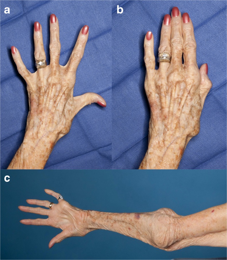
Fig. 4.
Postoperative appearance. Clinical photographs of the left hand and forearm taken on postoperative day 20. The patient has gained finger abduction (a) and adduction (b) indicating at least partial recovery of ulnar nerve function. c Clinical photograph taken from a posterior vantage point at 9 months follow-up. The fingers are actively abducted and the radius is dislocated anteriorly in this position. The olecranon can be seen prominently. There is no fluid collection at the incision site which has healed well

