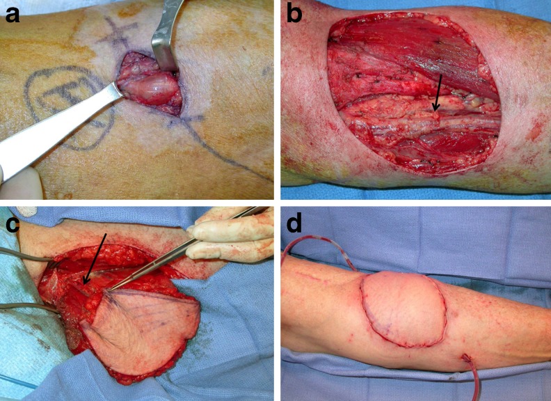
Fig. 2.
Patient 8 subsequently developed lesions suspicious for recurrence in the antecubital fossa and ring finger. These proved to be benign. a View of antecubital lesion (hand is to the right). b Defect following wide local excision. Ulnar artery (arrow). c A left free gracilis flap was harvested for soft tissue coverage. Flap pedicle (arrow). d Image of inset flap following end-to-end anastomosis to the ulnar artery

