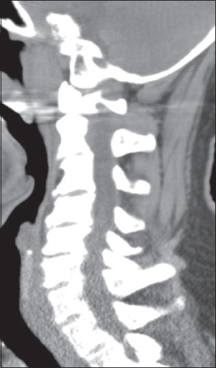
Figure 18.

Patient from Fig.17 Underwent CT in Conjunction with MR Documenting C5-C7 HPLL and Dorsolateral Shingling of the Laminae of C5, C6, C7 with Hyperlordosis. On the CT study, the ventral compression opposite the disc spaces of C4-5, C5-C6, and C6-7 was compatible with HPLL (non ossified but hypertrophied PLL). Note the direct visualization of the hyperlordosis and inward shingling of the lamina of C5, C6, C7
