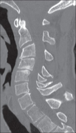
Figure 21.

Midline Sagittal 2D-CT Showing Marked Dorsolateral Shingling of the Laminae involving C4, C5, C6, C7. The patient's axial soft tissue window CT demonstrated marked cord compression from ventral multilevel HPLL and the patient underwent a posterior decompression/fusion
