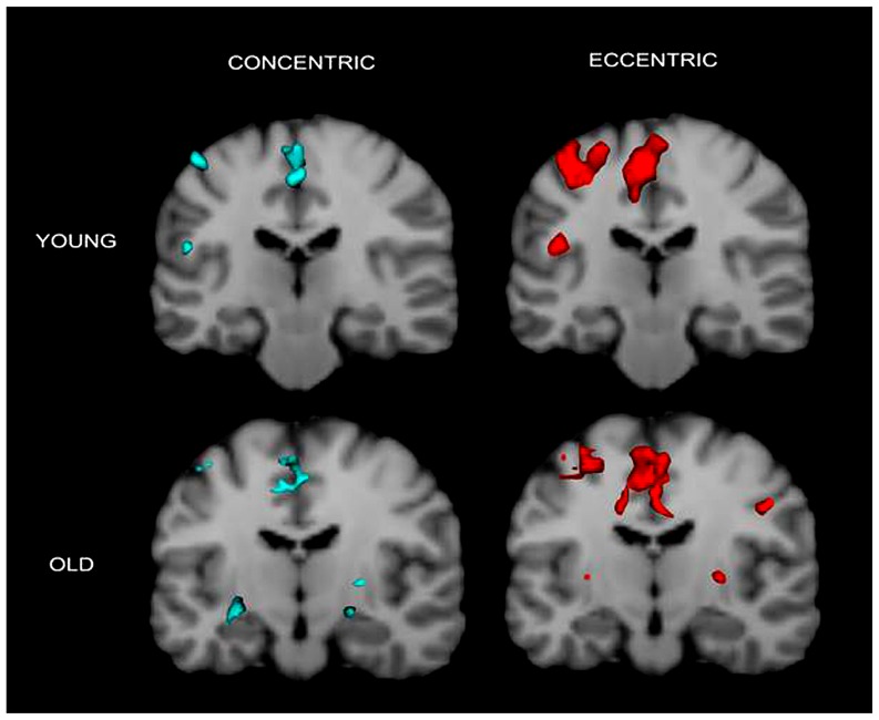
FIGURE 3.
Cronal view of group activation during the two types of contractions in the two groups. Again the figure shows that brain activation in young individuals are more localized in the contralateral (left) hemisphere but that in older adults are more distributed in the two hemispheres. Colored spots in the near-top central regions represent activated voxels in SMA and ACC.

