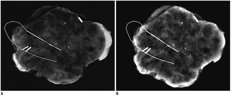
Fig. 4.
Specimen mammograms of ductal carcinoma in situ.
Mammography (B) was superior to mammography (A) for visualizing calcifications. However, number of calcifications was determined equally well using both mammography protocols. A. Image scanned by specimen mammography without application of "Mammogram enhancement ver. 2.0". B. Image scanned by specimen mammography with application of "Mammogram enhancement ver. 2.0".

