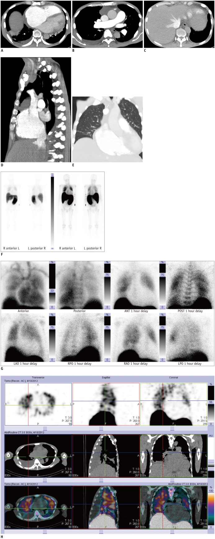
Fig. 1.
66-year-old man with primary myelofibrosis.
A-D. Chest CT images show contrast-filled dilated main pulmonary artery, dilated right atrium with retrograde opacification of inferior vena cava and hepatic veins due to tricuspid regurgitation. E. Chest CT (lung window-settings) did not show any lung parenchymal abnormality.
F. Tc-99m sulfur-colloid bone marrow scan image shows hepatosplenomegaly and moderately increased tracer activity in both hemithoracic regions. G. Tc-99m sulfur-colloid bone marrow scan (planar spot views) shows moderate degree of tracer uptake in diffuse pattern in both thoracic regions. Cardiac silhouette is enlarged. ANT = anterior, LAO = left anterior oblique, LPO = left posterior oblique, POST = posterior, RAO = right anterior oblique, RPO = right posterior oblique, Tc-99m = Technetium-99m
H. Single-photon emission computed tomography/CT images show pulmonary tracer (Technetium-99m sulfur colloid) activity in both lungs confirming pulmonary hematopoiesis.

