
Figure 7.
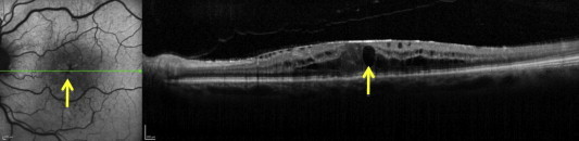
A diffuse area of hypoautofluorescence area involving the fovea and the surrounding area in an eye with cystic macular edema associated with central retinal vein occlusion. Arrow on the left side of the image is pointing toward the area of hypoautofluorescence and arrow on the right side is pointing toward cystic changes in the macula.