FIGURE 3.
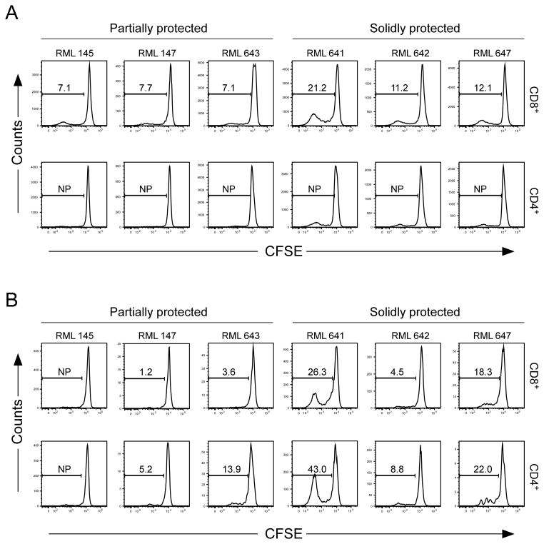
Chlamydial-specific T cell responses in boosted challenged macaques. CFSE labeled PBMC from boosted ocularly challenged macaques were incubated for five days with C. trachomatis strain A2497P+ EBs, A2497P+ SA prepared from infected BGMK cells, or with their respective negative controls; SPG or control SA. Cells were re-stimulated for six additional hours in the presence of anti-CD28 and CD49d and brefeldin A. Chlamydial antigen-specific T cell expansion was monitored by dilution of the CFSE proliferation marker using flow cytometry analysis. The flow analysis was performed on gated live CD3+ CD4+ and CD3+ CD8+ T cells. (A) CD8+ and CD4+ proliferation following incubation with A2497P+ SA. (B) CD8+ and CD4+ proliferation following incubation with A2497P+ EB. The numbers represent the percent of proliferation detected in PBMCs incubated with either chlamydial SA or EB normalized against respective negative control antigens. Note that CD8+ T cells of animals in the SP group (641, 642, and 647) proliferate more intensely and consistently in comparison to PP animals following stimulation with chlamydial SA.

