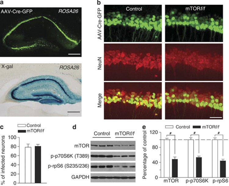
Figure 6.
AAV2-Cre-GFP-mediated deletion of mTOR in the hippocampus. (a) X-gal staining of hippocampus following intra-hippocampus microinjections of AAV2-Cre-GFP. AAV2-mediated Cre recombinase expression was labeled by LacZ (blue) when injected into Rosa26 reporter mice in the hippocampus. Scale bar: 0.5 mm. (b, c) Immunofluorescence staining for Neuronal Nuclei (NeuN; neuronal marker) and AAV2-Cre-GFP (green) in the hippocampus. Representative images (b) and summarized data (c) showed that AAV2-Cre-GFP infected ∼80% of all hippocampal CA1 pyramidal neurons in both control and mTORf/f mice (n=3 animals each group). Scale bar: 50 μm. (d and e) Representative (d) and summarized data (e) of western blots showed that intra-hippocampal microinjection of AAV2-Cre-GFP significantly decreased protein levels of mTOR, p-p70S6K (T389), and p-rpS6 (S235/236) in the hippocampus (#p<0.001; n=6 animals each group). Immunoreactivity was normalized to GAPDH and presented as the percentage of that of the control mice.

