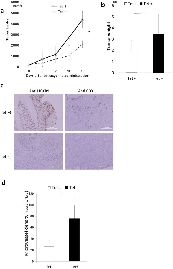
Figure 2.
Impact of HOXB9 expression on tumor xenografts. (a, b) Subcutaneous xenograft model of HT 29-T cells was established in Balb/c nu/nu mice (n = 8, each). Tumor burden (a) and tumor weight (b) were evaluated. (c) Immunohistochemical staining of paraffin-embedded xenografts obtained from HT29-T cancer cells. Samples were stained in anti-HOXB9 or anti-CD31. (d) Microvessel density of xenograft tumor was evaluated and measured in immunohistochemical staining by ant-CD31 antibody in xenografts (n = 8, each). Error bars are SDs. ☆ p < 0.05 and † p < 0.005 by U-test. Scale bars, 200 μm.

