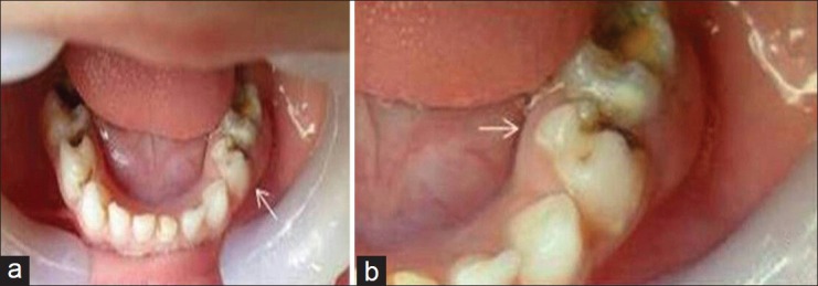
Figure 1.

(a) Clinical picture of the mandibular arch showing unilateral presence of fused macromolar L. Difference in shape can be appreciated by comparing with S, (b) Clinical crown shows extra mesio-lingual cusps of L resembling a rudimentary premolar with abnormal bulge and swelling in relation its attached gingival lingually.
