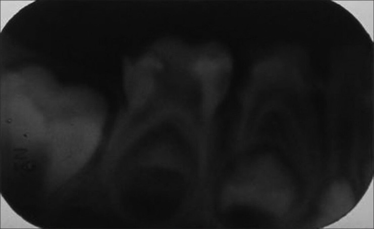
Figure 5.

The radiographic image of contralateral right mandibular molar is provided. It has only two roots with nor mal crown dimension which can also be seen in the clinical photograph. The dimension, presence of three roots, site of attachment of premolar and all side image of extracted tooth confirmed the fusion of mandibular first molar with supernumerary tooth.
