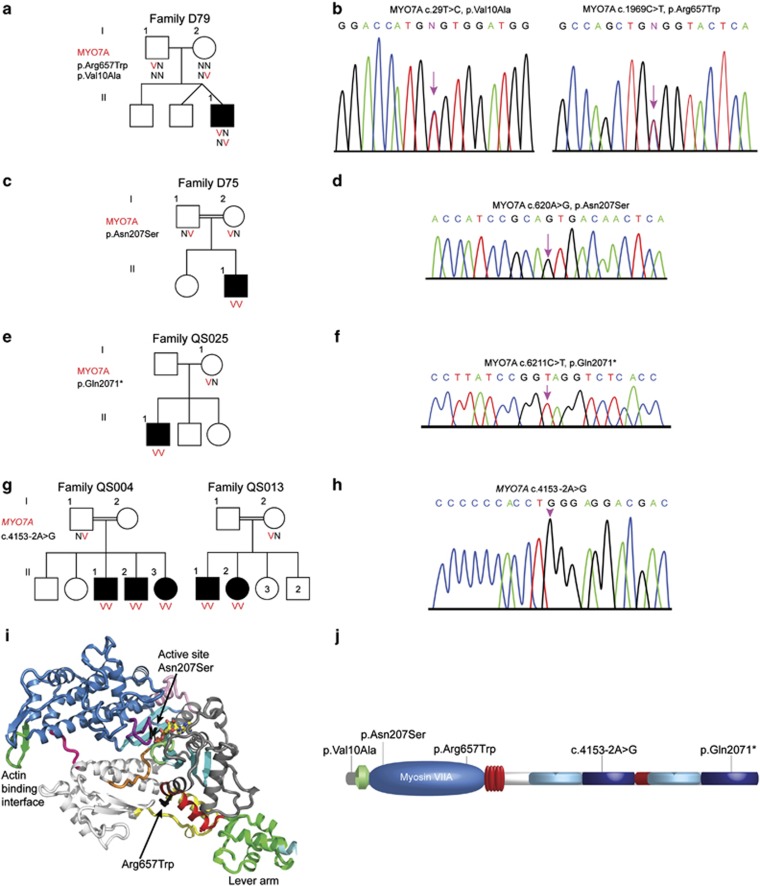
Figure 2.
Analysis of myosin VIIA mutations. (a) Pedigrees and segregation analysis of Family D79. (b) Partial sequences of MYO7A from a normal hearing and affected proband D79-II-1 demonstrating the c.29T>C and c.1969C>T mutations. (c) Pedigrees and segregation analysis of Family D75. (d) Partial sequences of MYO7A from an affected proband demonstrating the c.620A>G mutation. (e) Pedigrees and segregation analysis of Family QS025. (f) Chromatogram of the c.6211C>T mutation. (g) Pedigrees and segregation analysis of families QS004 and QS013. (h) Chromatogram of the c.4153-2A>G mutation. (i) Myosin VIIA mutations p.Asn207Ser and p.Arg657Trp are superimposed on a structural model of the myosin VIIa motor domain, based on the structure of myosin V bound to ATP.38 Note in black the location of the p.Asn207Ser mutation that may result in poor nucleotide binding and hydrolysis. The p.Arg657Trp mutation is likely to impair the communication between the active site and the lever arm as it is located at the interface between two connectors of the motor relay (yellow) and the SH1 helix (red) that controls the rotation of the lever arm. (j) Schematic representation of the myosin VIIA protein shows the location of p.Val10Ala, p.Arg657Trp, p.Asn207Ser, p.Gln2071* and c.4153-2A>G.

