FIG. 2.
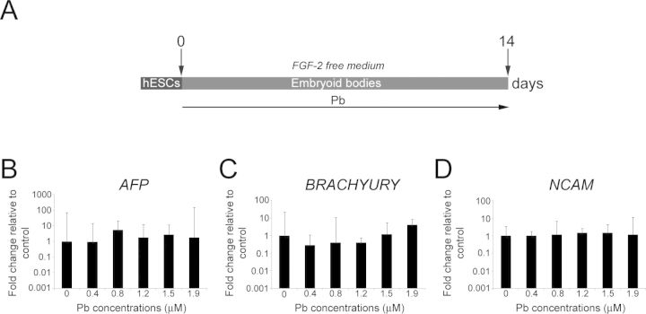
Pb exposure does not affect the potentials of hESCs to passively differentiate into cells of the three germ layers. (A) Schematic representation of the differentiation protocol and Pb exposure paradigm. (B) Quantitative RT-PCR expression analysis for endodermal (AFP), (C) mesodermal (BRACHYURY), and (D) ectodermal (NCAM) lineage markers in Pb-exposed hESCs after 2 weeks of differentiation in FGF-2-free medium. Embryoid bodies generated from control and Pb-exposed hESCs express markers of different germ layers. Histogram values represent means and error bars represent 95% interval confidences (p > 0.05; n = 3, one-way ANOVA).

