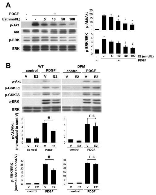
Figure 2. E2 inhibits PDGF-induced kinase activation in VSMC derived from WT but not DPM mice.
Western blotting and quantification of phosphorylated kinases in VSMC stimulated with PDGF (5 ng/ml) for 15 min with pretreatment of vehicle (V) or E2 for 30 min. (A) E2 inhibited phosphorylation of Akt and ERK in dose dependency in VSMC derived from WT mice (n=3). Quantification data is normalized to PDGF(−) vehicle-treated control. #P<0.05, *P<0.01. (B) E2-mediated inhibition of phosphorylation of Akt, GSK3α/β and ERK in VSMC derived from WT or DPM mice (n=4). #P<0.05.

