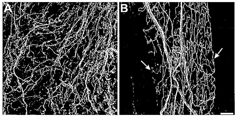
Figure 7.

Serotonin-like immunoreactivity in the foregut. A: Serotonin-immunoreactive fiber network in the anterior esophagus. B: Serotonin-immunoreactive fibers in the crop. The fibers were more uniform in diameter than those seen in the pharynx and esophagus. Although the patches did not correspond to any discernible underlying structures, they sometimes had clearly defined borders (arrows). No 5HTli cell bodies were detected in the foregut. Scale bar = 100 μm.
