Figure 5.
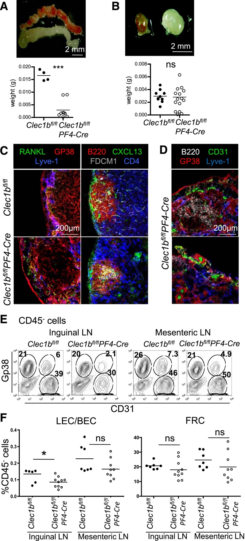
Impaired maintenance of the LNs in adults Clec1b−/−PF4-Cre. (A-B) Macroscopic appearance and weight of mesenteric (A) and inguinal (B) LNs from adult Clec1bfl/flPF4-Cre and Clec1bfl/fl control littermates. (C) Immunofluorescence staining of a section of adult LN from Clec1bfl/flPF4-Cre and Clec1bfl/fl control littermates stained for RANKL in green, Gp38/Podoplanin in red, and Lyve-1 in blue (first column) and B220 in red, CXCL13 in green, FDCM1 in gray, and CD4 in blue (second column), showing preserved LN structure in large inguinal Clec1bfl/flPF4-Cre LNs. (D) Immunofluorescence staining of section of fibrotic LNs from Clec1bfl/flPF4-Cre adult mice stained for B220 in white, CD31 in green, Gp38/Podoplanin in red, and Lyve-1 in blue. (E) Flow cytometric analysis of adult inguinal (first column) and mesenteric (second column) LN single-cell suspensions from Clec1bfl/fl control and Clec1bfl/flPF4-Cre littermates, stained with CD45, Gp38, and CD31. Percentages of CD31+Gp38− BEC, CD31+Gp38+ LEC, and CD31−Gp38+ FRC in the CD45− stromal fraction are indicated in the dot plots. (F) Quantification of the ratios of LEC:BEC (top chart) and percentages of FRC in the CD45− cell fraction (bottom chart) are shown. Unpaired Student t test, *P < .05, ***P < .001.

