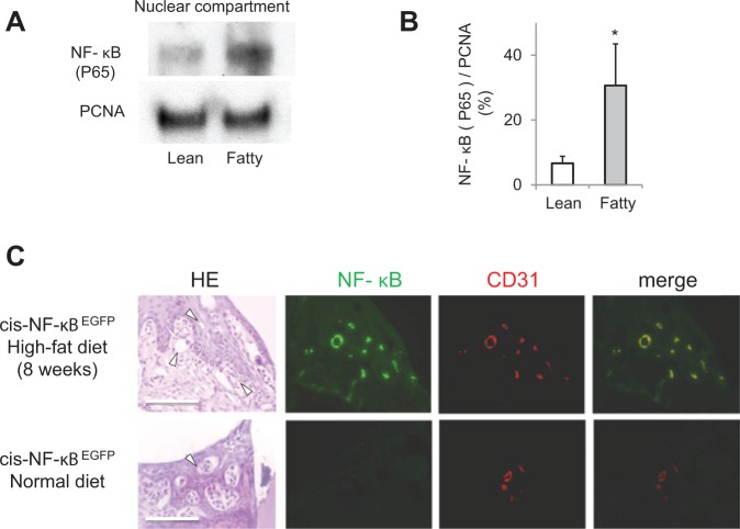
Figure 3.
Activation of NF-κB in the gingiva from insulin-resistant obese rodents. (A) Representative immunoblots of NF-κB (p65) from gingival nuclear proteins from Zucker rats. (B) Data from 3 experiments were normalized with PCNA and actin and quantified by densitometry. The p65 subunit of NF-κB was increased significantly in ZF rats. The data are expressed as mean ± SD. ZL vs. ZF, n = 6.*p < .05. (C) Representative immunohistological images in the mandibular periodontal tissue of cis-NF-κB-EGFP transgenic mice. The increased GFP expression in tissues demonstrated the activated NF-κB after rats consumed a high-fat diet for 2 mos. The GFP-positive areas were few in the normal-diet group. Immunostaining for CD31 and merged images with EGFP fluorescence was assessed by digital fluorescence microscopy. NF-κB activation was increased in the gingival endothelial cells from the mice consuming a HFD. Bar = 100 μm, n = 4 in each group.

