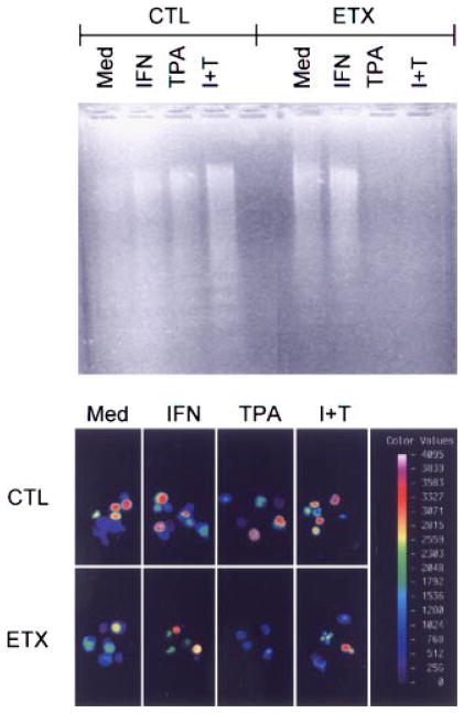
Fig. 2.
Effects of TPA and IFN-γ on DNA fragmentation. Neutrophils isolated from control (CTL) animals or 2 h after endotoxin (ETX) administration were cultured for 20 h with medium (Med), IFN-γ (I) and/or TPA (T). Upper panel: Soluble DNA extracts were analyzed on agarose gels and visualized using ethidium bromide. One representative gel from three separate experiments is shown. Lower panel: Cellular fluorescence intensity indicative of apoptosis was assessed using a Meridian ACAS 570. The color bar represents fluorescence on a four decade log scale. The color version of this figure is available online at www.interscience.wiley.com

