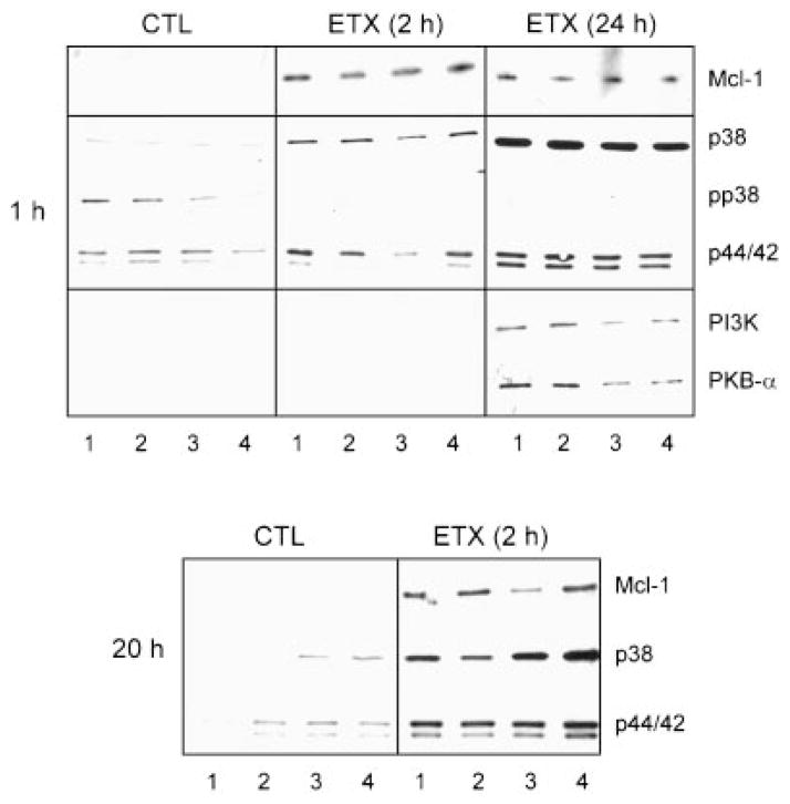
Fig. 3.
Effects of TPA and IFN-γ on Mcl-1, MAPK, PI3K, and PKB-α expression. Neutrophils isolated from control (CTL) animals or 2 or 24 h after endotoxin (ETX) administration were treated for 1 h (upper panel) or 20 h (lower panel) with medium control (lane 1), IFN-γ (lane 2), TPA (lane 3), or TPA + IFN-γ (lane 4). Extracts were analyzed by Western blotting as described in the Materials and Methods. One representative gel from three separate experiments is shown.

