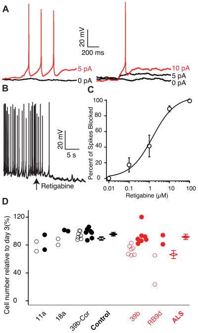
Figure 3. Retigabine Reduces Motor Neuron Excitability and Increases Survival.
(A) Rheobase measurements in a 39b Hb9::RFP-positive ALS-derived motor neuron in whole-cell patch clamp before (left) and after (right) the application of 10 μM retigabine (baseline rheobase 4.8 ± 1.5 pA vs post-retigabine rheobase 8.4 ± 2.2 pA; n=11; p<0.05, Wilcoxon signed rank test).
(B) Representative current clamp recording showing effect of 10 μM retigabine on membrane voltage and spontaneous firing (baseline Vm −60.4 ± 2.9 mV vs post-retigabine Vm −66.3 ± 3.6 mV, n=11; p=0.001, t-test). In (A–B), CNQX (15 μM), D-AP5 (20 μM), bicuculline (25 μM), and strychnine (2.5 μM) were added to the external solution.
(C) Dose response curve for retigabine on suppression of spontaneous action potentials in MEA recording and Hill plot fit of mean data from 39b (n=4) and RB9d (n=4) with EC50 1.5 ± 0.8 μM.
(D) Effect of vehicle (open circles) and 1μM retigabine (filled circles) treatment from days 14–28 of culture on the survival of Islet-positive, Tuj1-positive motor neurons measured at day 30 (total control n=11; total ALS n=9; F-test for effect of retigabine on all cells p=3.8×10⁻⁴; effect of retigabine in ALS motor neurons, red, 25.3% (SD 5.6; t-test p=6.4×10⁻⁵); effect of retigabine in control motor neurons, black, 6.1% (SD 5.1, p=0.23). Cell counts are from individual wells for four separate differentiations. See also Figure S4.

