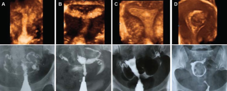
Fig 3.

Comparison of three-dimensional ultrasound and HSG imaging in cases of intrauterine lesions: A. Obliteration of the uterine cavity due to severe synechiae, B. Moderately extensive synechiae involving ½ of the uterine cavity, C. Endometrial polyp in the fundal area, D. Marked distortion and deformity of the uterine cavity caused by an intramural myoma bulging to the cavity.
