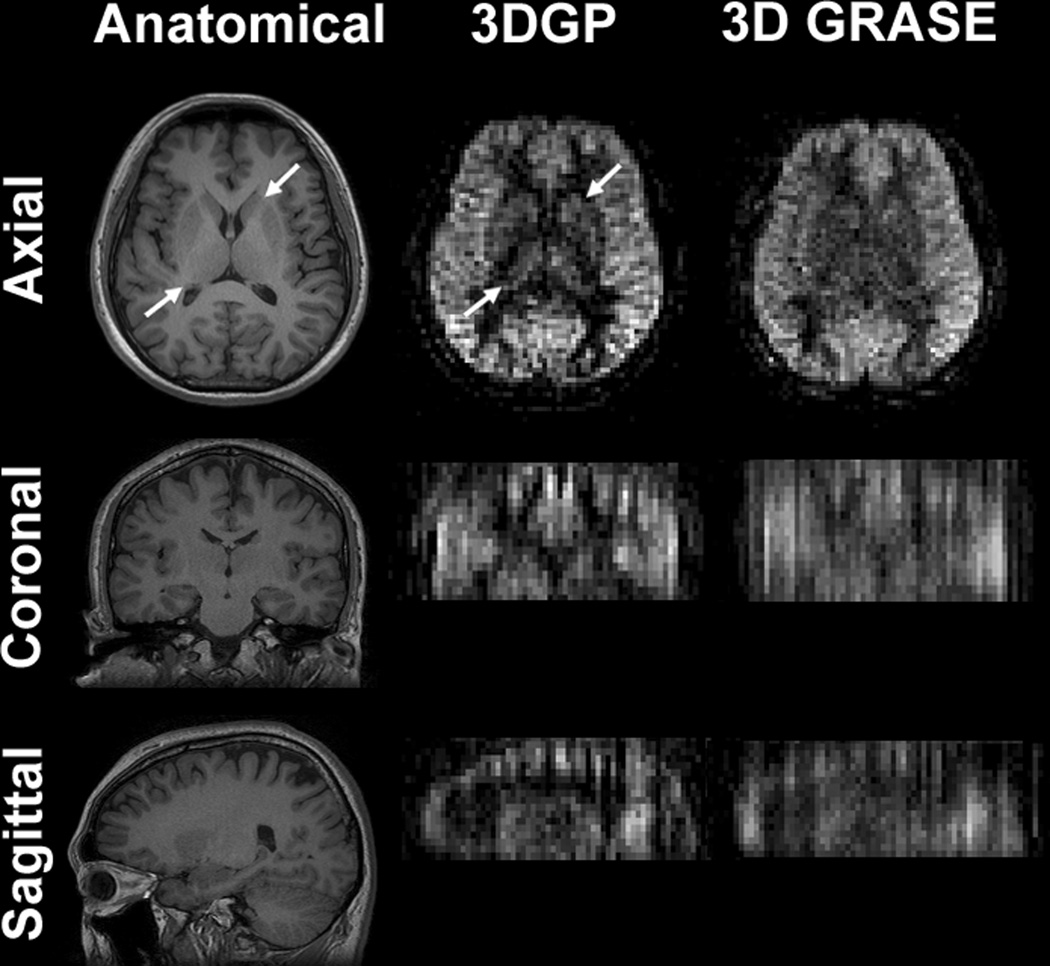
Figure 4.
Reformatted views of ASL images acquired in the axial plane. Severe blurring can be seen in the 3D GRASE images along the slice encoding direction. The blurring effect was significantly reduced in the 3DGP images. In-plane image details, such as thalamus and basal ganglia indicated by the arrows, can be easily identified in the 3DGP images whereas the same region was less recognizable in the 3D GRASE images.

