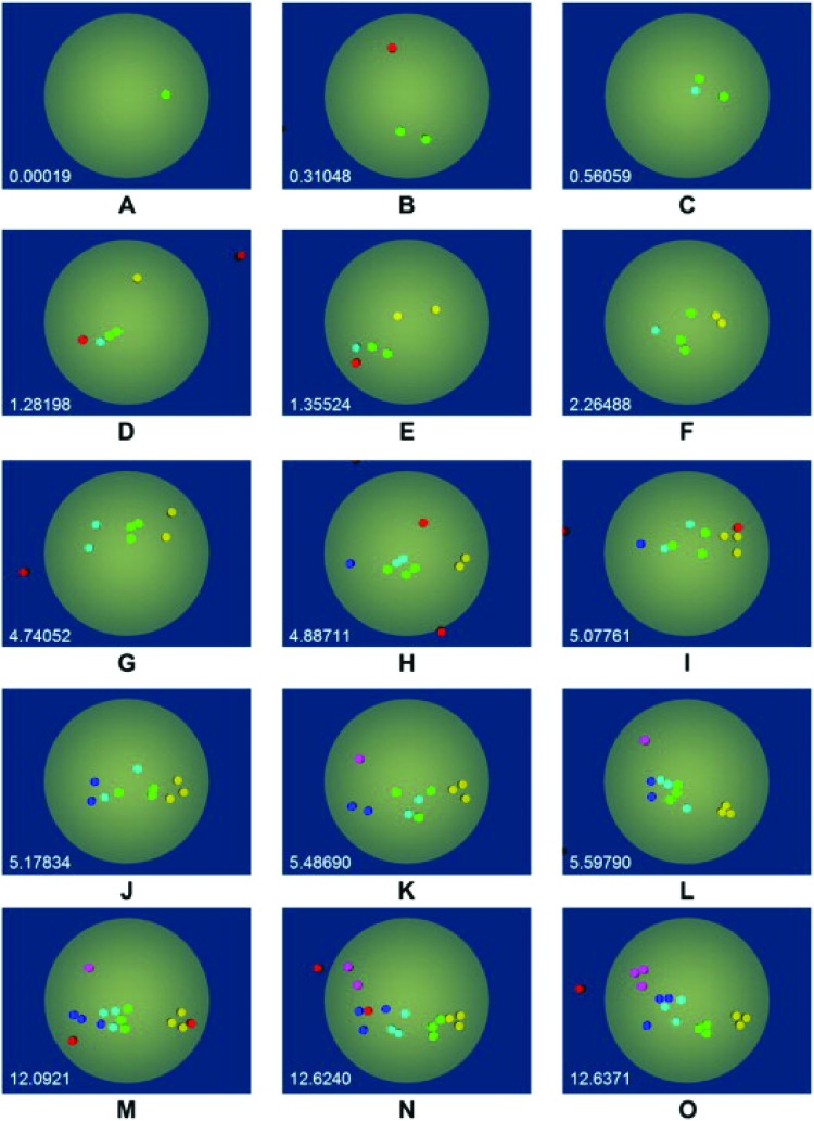
Fig. 7.
A simulation of the organization of TCR-gp120 bonds during the docking of HIV to the cell surface. Color coding indicates TCR that are bound to the same virus triskelion. The simulations indicate that after 12 s, the disorganized binding has minimized energy to lead to the formation of several well organized trimers. Figure reproduced from Ref. [57] with the permission of Elsevier.

