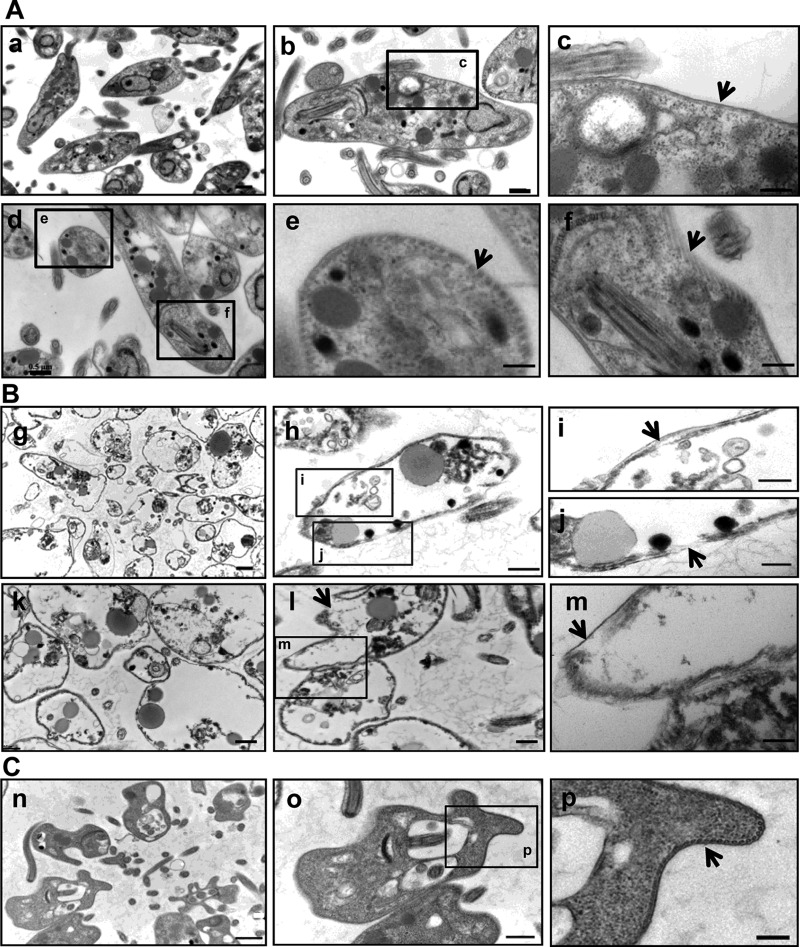
FIG 4.
Ultrastructural analysis of L. donovani promastigotes by transmission electron microscopy. (A) Untreated promastigotes with intact cytoplasmic organelles (a and d), intact plasma membrane (arrow) (b and c), or intact subpellicular microtubules (arrow) (e and f). (B) Promastigotes treated with compound 1, showing a high proportion of altered parasites (g and k), partial (h and i, arrow) or complete (m, arrow) detachment of the plasma membrane from the layer of subpellicular microtubules or complete disruption of the plasma membrane (j and l, arrow), or destruction of numerous intracellular organelles. (C) Compound 3-treated promastigotes, showing deformation of the plasma membrane (n and o) but no membrane rupture (p, arrow). Data are representative of two independent experiments. Scale bars represent 1 μm (a, g, and n) at magnification ×12,000 (12K), 0.5 μm (b, d, h, k, and o) at magnification 25K, or 0.2 μm (c, e, f, i, j, m, and p) at magnification 80K.

